
Articular Cartilage / Chondral Injuries
Articular Cartilage / Chondral Injuries
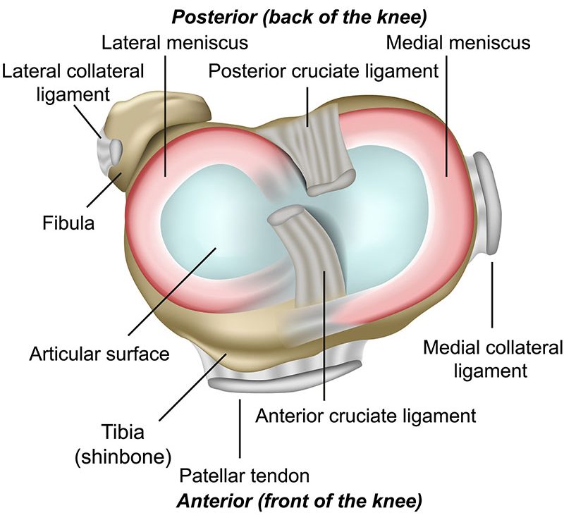
What is the articular cartilage?
The articular cartilage is a specialised protective layer over the bone ends within a joint. The term “chondral” is often used to refer to this cartilage. It has a role in spreading load and can be thought of as a low friction and smooth surface to allow the gliding motion to occur between the bones.
Injury
The articular surface can gradually thin and wear down with the ageing process and eventually lead to the problem of osteoarthritis. However, it can also be injured in more localised regions from a traumatic injury. This can either be in isolation or in conjunction with meniscal and ligament damage too.
Diagnosis
Most significant injuries are readily identified on modern MRI scanners and this is usually the initial investigation of choice. Simple X-Rays can be useful but are often unremarkable in the young patient with a traumatic chondral injury. The results of this MRI scan will then help to determine the appropriate course of action and guide expectations.
Treatment
Chondral injuries present us with one of the most challenging conditions in orthopaedics today. The problem being that this chondral cartilage has a poor inherent capacity to heal. Therefore, once damaged, problems of pain, stiffness, swelling and functional restrictions can develop. The extent of the damage and level of symptoms will determine then what options are available. Many can simply just be observed and monitored whilst others cause enough restriction that surgery needs to be considered. The surgical options can be complex and most need prolonged recovery times. The surgical options that might be discussed at consultation include: injections, chondroplasty, microfracture, osteochondral autograft transplantation (OATS), and occasionally more end-stage major options such as osteotomy and arthroplasty (a joint replacement).